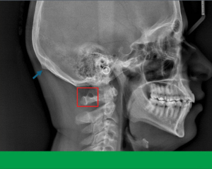
Orofacial Radiographic Manifestations in Patients with Sickle Cell Disease
 Original research in General Dentistry compared orofacial radiographs of patients with sickle cell disease (SCD) with those of a group of healthy controls. Generalized osteopenia, enlarged medullary spaces, thinning of the inferior mandibular border, and radiopaque lesions were significantly more frequent in patients with SCD.
 The findings of the study support the value of orofacial imaging for identifying signs of osteoporosis, reporting a high prevalence of enlarged medullary spaces in SCD patients. This feature is attributed to decreased bone density, bone marrow hypertrophy, and erythroblastic hyperplasia, all consequences of the high turnover of sickled erythrocytes.
Such findings demonstrate the potential of advanced imaging techniques to aid in the early detection and better management of bone alterations in SCD, providing a clearer understanding for dental professionals on how to approach and treat these patients more effectively.
Read more in the March/April issue of General Dentistry.
