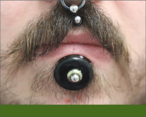
Large Piercing Hole Causes Imaging Artifact, Case Study Finds
 While it’s well known that jewelry can cause radiographic artifacts, a report in General Dentistry details a previously unreported type of anomaly produced when jewelry removal left a large soft tissue defect. The atypical radiolucency presented a diagnostic challenge until it was correlated with the piercing defect.
Case Report
A man who sought comprehensive care had multiple facial piercings, including a large piercing below the lower lip. The patient had undergone a 1-cm scalpel piercing of the skin under the lower lip and used progressively larger spacers to stretch the site over time. When the facial jewelry was removed prior to radiography, the aperture measured 28 mm and allowed a direct view of two mandibular incisors. The radiographic survey revealed a conspicuous horizontal, saucer-shaped radiolucency along the middle third of the mandibular incisors. The radiolucency, which mimicked the appearance of root resorption, severe dental caries or cervical abfraction, was ultimately attributed to the extensive aperture below the lower lip.
Summary
This case report describes a previously unrecognized intraoral radiographic presentation associated with a through-and-through piercing of the facial skin and labial mucosa. Clinicians should seek correlation of atypical radiographic presentations with soft tissue defects.
Read a synopsis of the full article here.
